
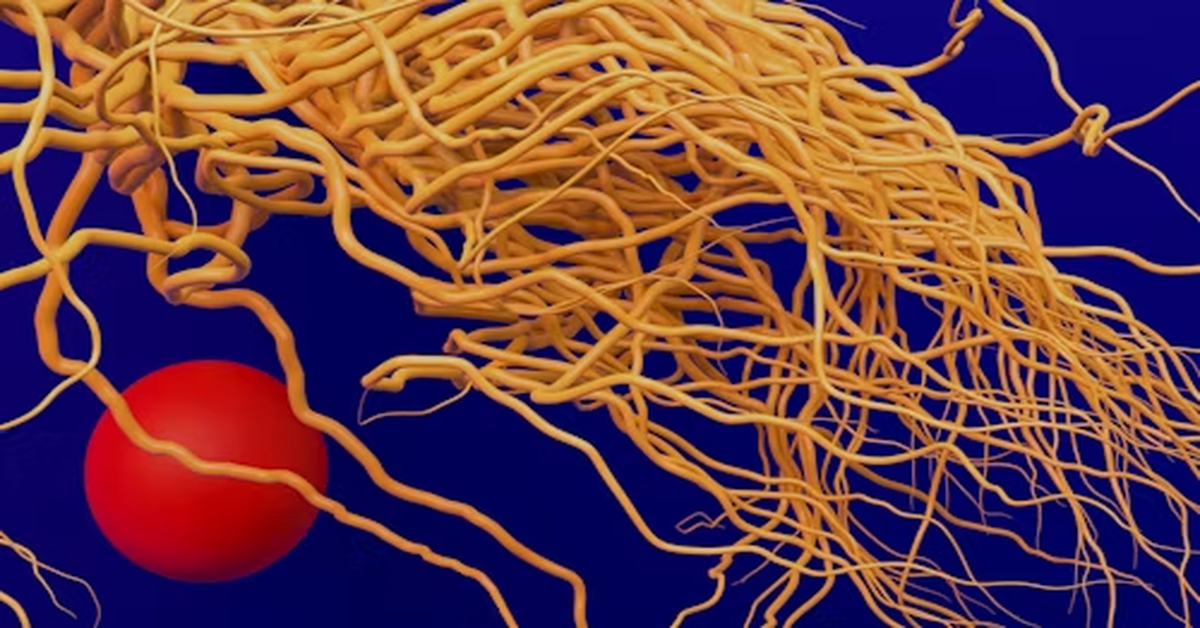
Gastroshiza is a congenital condition that affects the development of a baby’s abdominal wall during pregnancy. In this condition, the baby is born with the intestines protruding through an opening near the umbilical cord, usually to the right side. Unlike many other congenital defects, the organs in gastroshiza are not covered by a protective membrane, which makes them directly exposed to the amniotic fluid during pregnancy. This exposure can lead to irritation, inflammation, and sometimes damage to the intestines.
Though the correct medical term for this condition is gastroschisis, the term gastroshiza is often used colloquially or incorrectly to refer to the same defect. Regardless of the name used, understanding this condition is vital for expecting parents, healthcare professionals, and caregivers. Early diagnosis, appropriate prenatal care, and timely surgical intervention can lead to excellent outcomes in most cases.
In this article, we will discuss in detail the causes, risk factors, clinical presentation, diagnostic methods, surgical management, postoperative care, and long-term prognosis of gastroshiza. We will also include tables and sections to help you clearly understand every aspect of this condition.
What Is Gastroshiza?
Gastroshiza is a type of abdominal wall defect that occurs very early during fetal development—usually between the 4th and 8th week of pregnancy. During this time, the abdominal wall is supposed to close properly to keep the internal organs inside the body cavity. However, in babies with gastroshiza, this closure does not happen completely, leaving a small hole through which parts of the intestines (and sometimes other organs like the stomach or liver) can come out.
Unlike omphalocele—a similar but distinct condition—gastroshiza does not have a covering sac over the exposed organs. This difference is important because it increases the risk of infection, dehydration, and damage to the intestine.
Key Characteristics of Gastroshiza
| Feature | Description |
|---|---|
| Location | Typically occurs to the right of the umbilical cord |
| Protective membrane | Absent; intestines are directly exposed |
| Organs involved | Mainly intestines; sometimes stomach or liver |
| Associated abnormalities | Usually isolated; rarely associated with chromosomal defects |
| Treatment | Surgical closure soon after birth |
| Prognosis | Excellent with timely medical intervention |
Causes and Risk Factors
The exact cause of gastroshiza is not fully understood, but it is believed to result from a combination of genetic, environmental, and vascular factors. The defect happens when the developing abdominal wall fails to close completely, allowing parts of the intestine to protrude outside the body.
1. Genetic Factors
While gastroshiza is rarely inherited, certain genetic or chromosomal influences may contribute to its development. Unlike some other birth defects, it usually occurs as an isolated anomaly without other organ malformations.
2. Environmental Factors
Exposure to harmful substances during pregnancy—such as certain medications, alcohol, smoking, and recreational drugs—can interfere with fetal development and increase the risk of this defect.
3. Maternal Age
Studies have consistently shown that gastroshiza is more common in younger mothers, particularly those under 20 years of age. The reason is not entirely clear, but it may relate to vascular changes or nutritional deficiencies during early pregnancy.
4. Nutritional Deficiencies
Lack of adequate folic acid and other essential nutrients during pregnancy may interfere with normal tissue development, increasing the chance of defects in the abdominal wall.
5. Vascular Disruption
Some researchers believe that reduced blood flow to the developing abdominal wall could lead to tissue damage and failure of closure, resulting in gastroshiza.
6. Lifestyle and Environmental Exposure
Certain environmental toxins, pesticide exposure, or poor prenatal care environments may also play a role in triggering this condition.
Signs and Symptoms
Gastroshiza is usually evident at birth because the intestines are visibly protruding through a small opening in the abdominal wall. However, some signs can be identified before birth through prenatal ultrasounds.
Prenatal Signs:
- Abnormal findings on prenatal ultrasound showing loops of bowel outside the baby’s abdomen.
- Elevated levels of alpha-fetoprotein (AFP) in the mother’s blood test.
- Possible reduced fetal growth rate (intrauterine growth restriction).
Postnatal Signs:
- Visible protrusion of intestines through a small hole near the umbilicus.
- The exposed intestines appear thickened or swollen due to irritation from amniotic fluid.
- The baby may show signs of dehydration or difficulty regulating body temperature due to exposed organs.
- Feeding difficulties or delayed digestion after birth.
Diagnosis of Gastroshiza
Early and accurate diagnosis of gastroshiza plays a crucial role in planning prenatal care and postnatal surgery. The condition can be detected both before and after birth.
1. Prenatal Diagnosis
Most cases of gastroshiza are diagnosed during routine prenatal ultrasound scans, usually in the second trimester (around 18–20 weeks). The ultrasound shows loops of bowel floating outside the abdominal cavity, confirming the diagnosis.
Additionally, a maternal serum test called the triple or quadruple screen can show high levels of alpha-fetoprotein (AFP), suggesting an open abdominal wall defect. Once detected, the pregnancy is carefully monitored to ensure fetal growth and to plan for a safe delivery.
2. Postnatal Diagnosis
After birth, the diagnosis is visually apparent. A physical examination confirms the defect, and additional imaging (like X-rays or ultrasound) may be done to assess the extent of the herniation and check for any associated intestinal blockages or twisting.
Treatment and Surgical Management
Treatment for gastroshiza always involves surgical repair to place the intestines back inside the abdominal cavity and close the defect. The surgery must be done soon after birth to prevent infection, fluid loss, and intestinal damage.
Step-by-Step Overview of Treatment:
1. Immediate Postnatal Care
As soon as the baby is delivered, the exposed intestines are covered with a sterile, warm, and moist dressing to protect them from infection and drying. The baby is then placed in a temperature-controlled environment to prevent hypothermia.
2. Stabilization
The infant is stabilized by providing intravenous fluids, antibiotics, and oxygen. Feeding by mouth is avoided until the intestines are safely back inside and functioning normally.
3. Surgical Repair
There are two main surgical approaches:
a. Primary Closure:
If the exposed intestines are not too swollen, they are gently placed back into the abdomen, and the opening is closed in a single operation.
b. Staged Closure (Silo Method):
If the intestines are too large or swollen to fit back immediately, they are placed inside a special sterile pouch (called a silo bag). The intestines are gradually returned into the abdomen over several days, and then the opening is surgically closed.
4. Postoperative Care
After surgery, the baby is closely monitored in the neonatal intensive care unit (NICU). Feeding begins slowly through a tube until the intestines recover full function. The baby continues to receive fluids, nutrition, and antibiotics until they can feed normally.
Table: Primary vs Staged Closure in Gastroshiza Surgery
| Parameter | Primary Closure | Staged Closure (Silo) |
|---|---|---|
| Timing | Done immediately after birth | Gradual over several days |
| Use Case | Small defect, minimal swelling | Large defect, swollen intestines |
| Hospital Stay | Shorter | Longer |
| Risk of Complications | Slightly higher if pressure builds | Lower pressure, safer recovery |
| Outcome | Excellent with careful monitoring | Excellent, but longer process |
Possible Complications
Even though the prognosis of gastroshiza is generally very good, complications can occur—especially if the condition is not managed quickly or properly.
Short-Term Complications:
- Intestinal inflammation or damage due to exposure to amniotic fluid
- Infection from exposed tissue
- Dehydration and electrolyte imbalance
- Difficulty breathing if intestines take up too much space in the abdomen after closure
- Feeding intolerance or delayed bowel movement
Long-Term Complications:
- Short bowel syndrome (if part of the intestine is damaged or removed)
- Bowel obstruction due to scarring or adhesions
- Gastroesophageal reflux
- Slow growth or developmental delays (rare, but possible in premature infants)
With modern surgical and neonatal care, most babies recover completely and grow normally.
Prognosis and Long-Term Outlook
The prognosis for infants born with gastroshiza has improved dramatically over the last few decades. With advances in prenatal diagnosis, surgical techniques, and intensive neonatal care, survival rates now exceed 90–95% in most developed medical centers.
Most babies go on to live normal, healthy lives, although some may require additional medical follow-up for digestive or nutritional issues during infancy.
Factors that influence prognosis include:
- Size of the defect
- Amount of intestine exposed or damaged
- Timing and success of surgery
- Presence of other health complications
Prevention and Maternal Care
While gastroshiza cannot always be prevented, certain maternal health measures can reduce the risk.
Preventive Measures:
- Prenatal Vitamins: Regular intake of folic acid and multivitamins before and during pregnancy.
- Avoid Smoking and Alcohol: Both substances are linked to higher rates of congenital defects.
- Avoid Drug Use: Illicit drugs and some prescription medications can increase the risk.
- Healthy Diet: Adequate intake of protein, iron, and essential nutrients.
- Prenatal Checkups: Regular scans help in early detection and planning for safe delivery.
- Environmental Safety: Avoid exposure to harmful chemicals and pesticides.
Gastroshiza vs Omphalocele
Since both gastroshiza and omphalocele involve the abdominal wall, they are sometimes confused. However, they differ in many important ways.
| Feature | Gastroshiza | Omphalocele |
|---|---|---|
| Location | Right side of umbilical cord | Centered at the umbilical cord |
| Membrane covering | None | Has a sac covering the organs |
| Associated anomalies | Rare | Often associated with heart or chromosomal defects |
| Incidence | More common in young mothers | Can occur at any maternal age |
| Treatment | Surgical repair soon after birth | Surgical repair with more complex management |
Emotional and Psychological Support for Parents
Parents of infants with gastroshiza often experience anxiety, fear, and emotional exhaustion due to the uncertainty surrounding their baby’s condition. It’s essential for healthcare providers to offer emotional counseling, support groups, and clear communication about treatment progress and outcomes.
Encouraging parental involvement in care during the NICU stay—such as touching, talking to, and comforting their baby—can significantly improve bonding and emotional well-being for both parents and child.
Research and Future Outlook
Modern research is focusing on understanding the genetic and environmental causes of gastroshiza more deeply. Efforts are also being made to improve prenatal treatments, such as protecting the exposed intestines while the baby is still in the womb or developing safer methods for delivery and surgical repair.
The ultimate goal is to reduce complications, improve quality of life, and ensure every child born with gastroshiza has the best possible start.
Conclusion
Gastroshiza is a serious yet treatable congenital defect that requires immediate medical attention and careful postnatal care. With early prenatal detection, skilled surgical management, and advanced neonatal care, the prognosis for affected babies is excellent. Parents can take comfort in knowing that with the right medical team and timely intervention, most infants with gastroshiza grow up to live completely normal lives.
Understanding the condition—its causes, symptoms, and treatment options—empowers families and caregivers to make informed decisions and provide the best possible support for their child’s recovery and future health.
Frequently Asked Questions (FAQs)
1. What is gastroshiza?
Gastroshiza is a congenital abdominal wall defect in which the intestines protrude outside the baby’s body through a small hole near the umbilicus. It occurs during early fetal development.
2. How is gastroshiza diagnosed?
It is usually diagnosed during pregnancy through ultrasound and elevated maternal alpha-fetoprotein (AFP) levels, and confirmed after birth by physical examination.
3. Can gastroshiza be treated before birth?
Currently, surgical treatment is performed after birth. However, prenatal monitoring and planned delivery improve outcomes significantly.
4. What are the survival rates for babies with gastroshiza?
With modern surgical techniques and neonatal care, survival rates are above 90%, and most babies recover completely after treatment.
5. Can gastroshiza be prevented?
While not all cases can be prevented, healthy maternal habits—such as avoiding smoking, alcohol, and drugs, and ensuring proper nutrition—can lower the risk.